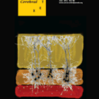
The cover image shows the connectivity between layer 5 pyramidal neurons in slice preparation of the medial prefrontal cortex (mPFC) of a developing mouse brain. Each cluster was filled with biocytin revealing cell soma location and dendritic branching patterns.
Developmental window for slow synapses & hyperconnectivity in Fragile X neocortex
From clinical observations made in functional brain imaging studies and post-mortem brain tissue from people with autism, a neurocognitive theory arose that connectivity was atypical between neurons in different brain regions in NeuroDevelopmental Disorders (NDDs) such as autism.
Alterations in the structure of dendrites and synaptic spines that connect neurons are observed in samples of brain tissue from both humans and genetic mouse models of many NDDs. We were curious to test whether the neurocognitive theory, based upon observations in the clinic, could apply to functional connections between neurons in living brain tissue.
Using a genetic mouse model for the mental retardation and autism spectrum disorder, Fragile X syndrome, we recorded the direct synaptic connections from small networks of neurons in brain slices made from the mouse prefrontal cortex (mPFC), since this brain region has specific roles in attention and executive function. Impairments in attentional behaviours and executive function are common to many NDDs, thus changes in the connectivity of prefrontal cortex neurons could underlie these behavioural changes.
Our experiments, recently published (Testa-Silva et al., 2011) reveal that there is hyperconnectivity between principal neurons in layer 5 mPFC in the mouse model for Fragile X syndrome. The chance of two neurons being synaptically connected was as high as 15% in some cases, compared with approximately 8% for control brain tissue. However, this change was transient during brain development– in older brains, no differences could be seen in neuron connectivity rates between both groups of mice.
Where there was a synaptic connection between two neurons, we also tested the kinetic and dynamic properties of the synapse. During the course of development, we were able to show that the dynamic responses of the synapses change: in 2-3 week old brain tissue, responses decreased in size when stimulated multiple times, a phenomenon known as a “depressing synapse‚. In contrast, brain tissue from 3-5 week old mice had synapse responses that increased in size, so called “facilitating synapses‚. This developmental change occurred in both groups of mice and is thought to be an essential property of the synaptic networks in mPFC to support working memory function in the brain.
However, there was a clear difference between the mice when it came to the temporal properties of synapses in these experiments. Using both experimental data and a computational model of the synapses, we measured the ability of the synapse to recover from being stimulated repeatedly, as could happen during information processing in the intact brain. In the mouse model for Fragile X, we found that the synapses were over 30% slower to recover following activation compared to those in control mouse brain. Changes of such a magnitude are likely to have significant effects on the processing and integration of neuronal information through these networks in the brain. And indeed, from collaboration with two researchers in Belgium and Germany who use computational modelling techniques, we were able to show that at the theoretical level, the combination of hyperconnectivity and slower synapses can alter the way that information is processed in this disorder.
Testa-Silva G, Loebel A, Giugliano M, de Kock CP, Mansvelder HD, Meredith RM (2012). Hyperconnectivity and slow synapses duringearly development of Medial Prefrontal Cortex in a mouse model for mental retardation and autism. Cereb. Cortex (2012) 22 (6): 1333-1342.
Highlighted on SFARI autism site : http://sfari.org/news-and-opinion/in-brief/2011/molecular-mechanisms-fragile-x-brains-have-altered-synapses